Radiology
To make an appointment from a physician referral or for more information, please call South Central’s Radiology Department at 601-426-4090.
FibroScan
For those who are battling with symptoms of liver disease, FibroScan® is an FDA approved technology that offers a quick and painless approach to liver health. Your physician may recommend a FibroScan® test if you have one of the following chronic liver conditions:
- Hepatitis B
- Hepatitis C
- Alcoholic Liver Disease
- Non-Alcoholic Steatohepatitis (NASH)
- Autoimmune Hepatitis
- Genetic Diseases such as Hemochromatosis and Wilson’s Disease
- Cirrhosis
Genius™ 3D Mammography™
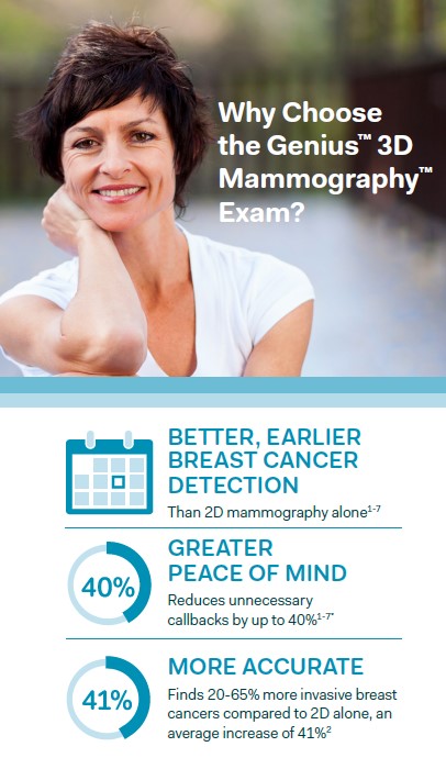 Utilizing advanced breast tomosynthesis technology, Genius exams are clinically proven to significantly increase the detection of breast cancers, and also decreasing the number of women asked to return for additional testing. In conventional 2D Mammography, overlapping tissue is a leading reason why small breast cancers may be missed and normal tissue may appear abnormal, leading to unnecessary callbacks. A Genius exam includes a three-dimensional method of imaging that can greatly reduce the tissue overlap effect.
A Genius exam includes both 2D images and tomosynthesis scans. During the tomosynthesis-DIMENSIONAL portion of the exam, an X-ray arm sweeps in a slight arc over the breast, taking multiple images. A computer then converts the images into a stack of thin layers, allowing the radiologist to review the breast tissue one layer at a time. A Genius exam requires no additional compression and takes just a few seconds longer than a conventional 2D breast cancer screening exam.
The Genius™ 3D Mammography™ (a.k.a. Genius™ exam) is acquired on the Hologic® 3D Mammography™ system and consists of a 2D and 3D™ image set, where the 2D image can be either an acquired 2D image or a 2D image generated from the 3D™ image set. The Genius™ exam is only available on the Hologic 3D Mammography™ system.
Our Genius™ 3D Mammography™ exam, available on the 3Dimensions™ and Selenia® Dimensions® systems from Hologic, are revolutionizing how breast cancer is detected by providing a better option for women of all breast densities compared to 2D alone. Researchers have found that:
- The Genius™ 3D Mammography™ exam finds 20-65% more invasive breast cancers compared to 2D alone, an average increase of 41%.
- Only the Genius™ 3D Mammography™ exam is FDA approved as superior for women with dense breasts compared to 2D alone.
- The Genius™ 3D Mammography™ exam reduces callbacks by up to 40% compared to 2D alone.
If you would like to schedule a Genius 3D Mammography™ exam, or have questions about this important breast health procedure, please contact South Central Breast Care Center at 601-426-4090.
Invenia™ ABUS – Dense Breast Screening
About 40% of women have dense breast tissue. And for these women, mammograms alone may not be enough to find breast cancer. South Central Regional Medical Center now offers Invenia™ ABUS, the only breast cancer screening technology FDA-approved for detection in women with dense breast tissue.
What does Breast Density mean? Breasts are made of fat and breast tissue. Some women have more fat than breast tissue while others have more breast tissue than fat. When there is more breast tissue the breast is considered dense. On a mammogram dense tissue looks white. Since masses or lumps also appear white on a mammogram, a suspicious lump may be masked by the dense breast tissue.
Dense breast tissue is also linked with an increase in the risk of developing breast cancer. Women with extremely dense breast tissue have a 4 to 6 times greater risk of developing breast cancer than women who do not have dense breast tissue. Invenia™ ABUS helps physicians look differently at dense breast tissue.
Breast density is determined by the radiologist who reads your mammogram and classifies the density into one of four categories. Your doctor will tell you if you have dense breasts based on your mammogram’s classification on the density scale.
DXA
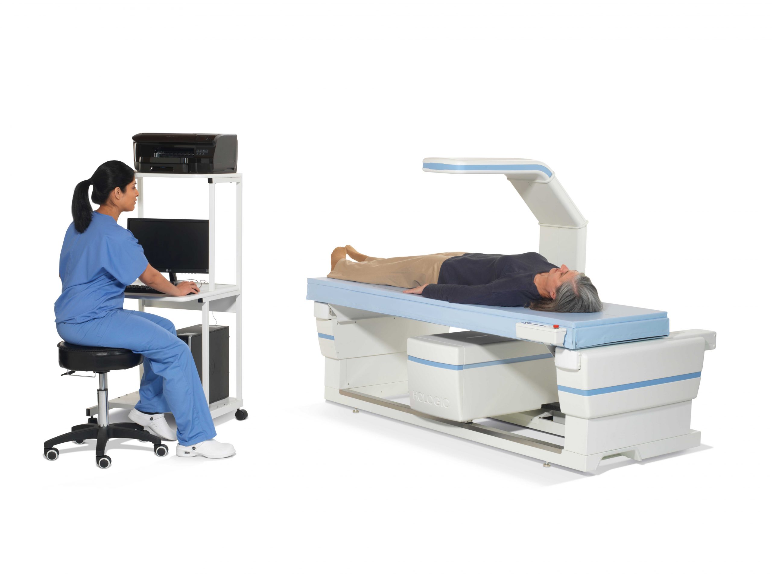 South Central’s Radiology Department has expanded its health and wellness services available to men and women with the addition of the Horizon® DXA system from Hologic®. The imaging technology of the Horizon DXA system provides superb image quality with advanced diagnostic tools to support early detection and treatment of osteoporosis.
Osteoporosis is a growing healthcare crisis affecting millions of women and men worldwide. 77% of American women with osteoporosis are undiagnosed (and therefore untreated). Fortunately, osteoporosis is detectable and treatable and testing is safe and non-invasive. The Horizon® DXA system at SCRMC enables new dimensions in care, including greater insights into biomechanical strength, BMD measurements of the entire skeleton, and many other clinical applications.
MRI
The MRI suite at SCRMC incorporates state of the art diagnostic imaging technology in a personalized healing environment. Our staff of board certified and state licensed Radiologists and Radiologic Technologists ensure that your MRI exam is performed and interpreted using the latest imaging techniques. It is our goal to offer a comfortable, anxiety free environment to our patients while providing exceptional imaging. We are proud of the fact that our MRI department is accredited and recognized by the American College of Radiology. We are pleased to offer ACR accredited MRI Breast Imaging. We also offer MRI Prostate Imaging with CAD.
We are also pleased to offer MRI Breast imaging as part of our commitment to provide a wide range of diagnostic imaging services to our patients.
PET/CT
South Central Regional Medical Center has the capability to provide the latest technological advancement in PET imaging, PET/CT (Hybrid/Fusion Imaging). The PET/CT (Hybrid/Fusion Imaging) allows for the physiologic assessment of the human body with an anatomical overlay.
South Central has acquired state-of-the-art PET equipment that has the capability of performing both PET and CT examinations simultaneously. This newest innovation allows for PET imaging to more accurately pinpoint abnormalities in the human body, thereby allowing physicians to diagnostically evaluate conditions and diseases with the latest technology.
Nuclear Medicine
Nuclear medicine is a specialized area of radiology that uses very small amounts of radioactive materials to examine organ function and structure. This branch of radiology is often used to help diagnose and treat abnormalities very early in the progression of a disease.
Because X-rays pass through soft tissue, such as intestines, muscles, and blood vessels, these tissues are difficult to visualize on a standard X-ray, unless a contrast agent is used. This allows the tissue to be seen more clearly. Nuclear imaging is used to study organ and tissue function.
South Central Regional Medical Center’s Nuclear Medicine department focuses on the following areas:
- Renal scans
- Thyroid scans
- Bone scans
- Gallium scans
- Heart scans
- Brain scans
- Breast scans
- Parathyroid scans
- Liver/Spleen scans
- Gallbladder scans
- Gastric scans
- Lung scans
- GI Bleed scans
64 Slice CT (Computerized Tomography)
We offer computerized tomography services (CT) using the latest technology. We obtain your images using GE VCT 64 slice scanners. We have two scanners to help minimize wait for our patients. Both of our scanners are equipped with the latest in radiation dose reduction software. We also offer fluoroscopic guided biopsy and cardiac imaging on our scanners.
We are American College of Radiology (ACR) accredited on both of our CT scanners. We are also proud supporters of Imaging Gently and are accredited by the ACR in pediatric imaging as recognized by The Alliance for Radiation Safety in Pediatric Imaging.